

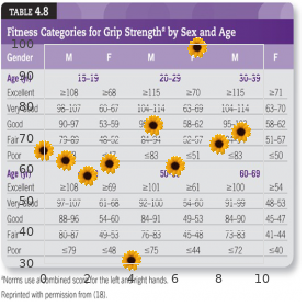
State University of New York College at New Paltz. X. Fabio, MD: "Buy online Cephalexin cheap no RX - Discount online Cephalexin OTC".
Ligatures used range from electrical cords buy cephalexin in united states online antibiotics for acne that don't cause yeast infections, neckties buy discount cephalexin online antibiotic cipro, ropes generic 500 mg cephalexin antimicrobial vapor barrier, and telephone cords to sheets and hose. The appearance of a ligature mark on the neck is subject to considerable variation, depending on the nature of the ligature, the amount of the resistance offered by the victim, and the amount of force used by the assailant. The ligature mark might be faint, barely visible, or absent in young children or incapacitated adults, especially if the ligature is soft, e. If a thin tough ligature is used, there will be a very prominent deep mark encir- cling the neck. In ligature strangulation, in contrast to hangings, the ligature mark usu- ally encircles the neck in a horizontal plane often overlying the larynx or upper trachea (Figure 8. There might be a break in the furrow, however, usually in the back of the neck, where a hand has grasped the ligature and tightened it at this point. Aside from the ligature mark, abrasions and contusions of the skin of the neck are usually not present. If there is more than one loop of the ligature around the neck, there could be bruising of the skin if the ligature pinches the skin between two loops. Injury to the internal structures of the neck is the exception rather than the rule. In a study of 48 ligature strangulation deaths, fractures of the hyoid or thyroid cartilage were present in only six cases (12. Four of the victims had fractures of both the thyroid and 260 Forensic Pathology Figure 8. In such cases, the victim usually ties a ligature tightly around the neck (Figure 8. Because some individuals remain conscious for 10–15 sec after complete occlusion of the cartoid arter- ies, they have sufficient time to tie at least one, if not more, knots. Instead of tying a knot, some individuals tightly wrap a ligature several times around the neck, securing it in place by the overlapping loops. Other individuals use a tourniquet method: a ligature is loosely wrapped around the neck, knotted, and then tightened by a stick inserted beneath the ligature and twisted mul- tiple times. Clothing or the individual’s own weight on the stick holds it in place, maintaining the “tourniquet. It is seen when a tie, scarf, shirt, or other article of clothing gets entangled in a moving machine. Isadora Duncan, the dancer, died of accidental strangulation when a scarf she was wearing became entangled in an automobile wheel. In infants, the elderly, and decomposing bodies, there can be pseudo-ligature marks that suggest strangulation. In 262 Forensic Pathology infants, pale crease marks between overlapping folds (rolls) of neck skin can be mistaken for ligature marks. This can be further complicated in newborns by the presence of petechiae, which can be normally present in newborns delivered vaginally because of compression of the chest. A similar picture of pseudo-ligature marks can be seen in the elderly — pale crease marks caused by overlapping rolls of skin, petechiae of the sclerae and conjunctivae (caused by cardiac failure) — with the addition of retro- pharyngeal hemorrhage. Petechiae are often seen in acute cardiac failure, not uncommonly the cause or mechanism of death in elderly individuals. The case that comes to mind is that of an elderly woman found dead in bed with a pillow propping up her head, such that her chin was against her chest. When the body was examined at the medical examiner’s office, there was a horizontal mark across the front of her neck that simulated a ligature. The face above the mark was congested and there were fine petechiae of the sclerae, conjunctivae, and periorbital skin, as well as retropharyngeal hemorrhage. At the time the body was initially autopsied, a full account of the circumstances and scene surrounding the death was unknown. For example, it was not known that the head had been propped up, that there was a long history of cardiac failure, and that the woman died in a room with no windows and with a single entrance that was blocked by a bed in which her bedridden husband slept. The “ligature mark” was just an artifact — a crease caused by the positioning of the head. Pseudo- ligature marks can also be seen in decomposing bodies with tight collars or other clothing around the neck. The body, as it decomposes, swells around the tight-fitting garment, which produces a deep furrow that simulates a ligature mark. In victims of homicidal ligature strangulation, hair is often found clutched in the hands. A control sample of the victim’s hair should be obtained for comparison, because the hair found in the hands almost invariably turns out to be that of the victim. Fingernail scrapings or cuttings (the latter are preferred) should be taken to look for tissue of the perpetrator under the nails. Unfortunately, unlike in fiction, such scrapings or cuttings have traditionally been of little help, with foreign tissue rarely identified. Manual Strangulation Manual strangulation is produced by pressure of the hand, forearm, or other limb against the neck, compressing the internal structures of the neck. The mechanism of death is occlusion of the blood vessels supplying blood to the Asphyxia 263 brain i. In the authors’ experience, it is the second most common method of homicidal asphyxia. In a study by DiMaio of 41 deaths caused by manual strangulation, females predominated, with the ratio of females to males 1. One cannot commit suicide by manual strangulation because, as soon as consciousness is lost, pressure is released and consciousness regained. Occasionally, it is claimed that the death of a healthy individual ascribed to manual strangulation is unintentional and caused by a vasovagal reaction (reflex cardiac death) brought on by touching, grasping, or striking the neck. The mech- anism of death in such a case would be an arrhythmia produced by stimu- lation of the carotid sinuses. The carotid sinus is a focal area of enlargement of the common carotid artery where it bifurcates into the external and inter- nal carotid arteries. Compression or stimulation of the carotid sinuses causes an increase in blood pressure in these sinuses with resultant slowing of the heart rate (bradycardia), dilatation of blood vessels (vasodilation), and a fall in blood pressure. Pressure on the common carotid artery below the sinuses reduces the blood pressure within the sinus by reducing the amount of blood flowing into it. This mimics hypotension or decreased blood supply from hemorrhage or shock, causing the heart to beat faster (tachycardia), the blood vessels to constrict (vasoconstriction), and a rise in blood pressure. This explains the fact that, while in most cases of manual strangulation there is bradycardia, vasodilation, and fall in blood pressure, in some cases, if the hands are lower down on the neck, there might instead be tachycardia, vasoconstriction, and a rise in blood pressure. In normal individuals, pressure on the carotid sinus causes minimal effects with a decrease in heart rate of less than six beats per minute and only a slight reduction in blood pressure (less than 10 mm Hg). In such individuals, there is slowing of the heart and cardiac arrhyth- mias ranging from ventricular arrhythmias to cardiac stand-still and hypotension. There are cases reported in which turning of the neck in varying positions or a high or tight collar has produced dizziness and fainting. Di Maio found petechiae present in the conjuctivae or the sclerae in 89% of his cases.
The reactivation of these fetal genes buy cephalexin american express antibiotics for hotspots on dogs, the so-called fetal gene program cephalexin 250mg with visa tween 80 antimicrobial, also is accompanied by decreased expression of a number of genes that are normally expressed in the adult heart buy genuine cephalexin on line best antibiotics for sinus infection doxycycline. As discussed later, activation of the fetal gene program may contribute to the contractile dysfunction that develops in the failing myocyte. These stimuli occur both locally within the myocardium, where they exert autocrine/paracrine effects, and systemically, where they exert endocrine effects. The early stage of cardiac myocyte hypertrophy is characterized morphologically by increases in the number of myofibrils and mitochondria, as well as enlargement of mitochondria and nuclei. At this stage, the cardiac myocytes are larger than normal, but with preservation of cellular organization. As hypertrophy continues, there is an increase in the number of mitochondria, as well as the addition of new contractile elements in localized areas of the cell. Cells subjected to longstanding hypertrophy show more obvious disruptions in cellular organization, such as extremely enlarged nuclei with highly lobulated membranes, accompanied by the displacement of adjacent myofibrils with loss of the normal registration of the Z-bands. The late stage of hypertrophy is characterized by loss of contractile elements (myocytolysis) with marked disruption of Z-bands and severe disruption of the normal parallel arrangement of the sarcomeres, accompanied by dilation and increased tortuosity of T tubules. Alterations in Excitation-Contraction Coupling As discussed in Chapter 22, excitation-contraction coupling refers to the cascade of biologic events that begins with the cardiac action potential and ends with myocyte contraction and relaxation (see Fig. Impaired contraction and relaxation of the failing heart is most prominent at high heart rates, which results in a depressed force-frequency relationship. This has been demonstrated both in isolated strips of human myocardium and in clinical observations of patients (Fig. Normally, higher contraction frequency 2+ increases cardiac performance because of a frequency-dependent augmentation of intracellular Ca transients. By contrast, in the failing myocardium, a decline in force generation is seen with higher heart 2+ 2+ rates that is secondary to a decrease in amplitude of intracellular Ca , a prolonged decline of the Ca 2+ transient, and increased levels of diastolic calcium. A, Relationship between stimulation frequency and force generation of isolated muscle strip preparations from nonfailing and failing human hearts. In nonfailing myocardium, contractile force increases up to a stimulation rate of approximately 2. Heart rate was changed by temporary pacing during cardiac catheterization, and cardiac output was measured by thermodilution. Ca2+ handling and sarcoplasmic reticulum Ca2+ content in isolated failing and nonfailing human myocardium. Influence of the force-frequency relationship on haemodynamics and left ventricular function in patients with non-failing hearts and in patients with dilated cardiomyopathy. This observation has led to the suggestion that the increase in contractile function following treatment with beta blockers is secondary to RyR stabilization. Action Potential Duration and Sodium Handling Several factors contribute to the prolongation of the action potential duration, which is a ubiquitous 27 finding in failing hearts. As discussed in Chapter 22, the voltage-gated Na channels are activated on + depolarization of the cell membrane, leading to rapid influx of Na that is responsible for the fast upstroke + of the action potential (see eFig. Under normal conditions, Na channels inactivate a few + milliseconds after depolarization. High levels of intracellular + Na also may lead to cellular acidosis secondary to increased sodium-proton exchange activity. Under normal conditions, Na channels inactivate a few milliseconds after + + depolarization and net Na influx is small. Persistent + + opening of Na channels in the myocyte from a failing heart results in a larger Na influx relative to that for the myocyte from the normal heart. Myocardial gene expression in dilated cardiomyopathy treated with beta blocking agents. Changes in myosin light chain isoforms have been observed in the atria and ventricles of patients whose hearts have been subjected to mechanical overload. Abnormalities in Cytoskeletal Proteins The cytoskeleton of cardiac myocytes consists of actin, the intermediate filament desmin, the sarcomeric protein titin (see Chapter 22), and alpha- and beta-tubulin, which form the microtubules by polymerization. Vinculin, talin, dystrophin, and spectrin constitute a separate group of membrane- associated proteins. Loss of integrity of the cytoskeleton and its linkage of the sarcomere to the sarcolemma and extracellular matrix would be expected to lead to contractile dysfunction at the myocyte level, as well as at the myocardial level. The binding of beta-arrestins to the cytoplasmic tail of the beta receptor not only uncouples the receptor from heterotrimeric G proteins, but also targets the receptor for internalization in clathrin-coated vesicles. Although this internalization fosters receptor dephosphorylation and serves as a prelude to recycling the beta receptor to the surface for reactivation, at some point receptor entry via endocytosis is not followed by recycling, but rather leads to receptor trafficking to lysosomes and receptor degradation. However, by reducing energy expenditure of the energy-starved myocardium and protecting the myocyte from the deleterious effects of sustained adrenergic stimulation, this adaptive response is beneficial. Alterations in the Myocardium The changes that occur in failing myocardium may be categorized broadly into those that occur in the volume of cardiac myocytes and those that occur in the volume and composition of the extracellular matrix. Although necrosis initially was thought to be a “passive” form of cell death, emerging evidence indicates 33 that necrotic cell death also is “regulated. Dysfunction of the plasma membrane in necrotic cells leads to cell swelling and rupture. In the heart, increased plasma membrane permeability allows Ca to leak into the cell, exposing the contractile proteins to very high concentrations of this activator, which in turn initiates extreme interactions between the myofilaments (contraction bands), further contributing to disruption of the cellular membrane. Necrotic myocyte death occurs in ischemic heart disease, myocardial injury, toxin exposure (e. The final result is a fibrotic scar, which may alter the structural and 34 functional properties of the myocardium. Information about regulated signaling in necrosis is currently limited to two pathways. The intrinsic pathway is activated by diverse biologic, chemical, and physical stimuli. These death signals trigger the release of apoptogens from the mitochondria into the cytosol, including cytochrome c, which triggers the formation of a second multiprotein complex, the apoptosome, in which procaspase-9 undergoes activation. Downstream caspases cleave several hundred cellular proteins to bring about the apoptotic death of the cell. Apoptosis, or programmed cell death, is an evolutionarily conserved process that allows multicellular organisms to selectively remove cells through a highly regulated program of cell suicide. In addition, connections between the pathways amplify signals, increasing the efficiency of killing. Apoptosis plays important roles in development and in postnatal life, when it is critical for tissue homeostasis and surveillance for damaged or transformed cells. In contrast with the cell swelling that characterizes necrosis, during apoptosis the cell shrinks and eventually breaks up into small, membrane-surrounded fragments. The latter often contain bits of condensed chromatin referred to as apoptotic bodies. Maintenance of plasma membrane integrity until late in the apoptotic process allows the dying cell to be engulfed by macrophages, which prevents the release of the reactive intracellular contents, thereby preventing an inflammatory reaction. Autophagy refers to the homeostatic cellular process of sequestering organelles, proteins, and lipids in a double-membrane vesicle inside the cell (autophagosome), where the contents are subsequently delivered to the lysosome for degradation. Unlike necrosis and apoptosis, autophagy is primarily a survival mechanism that regulates the quality and abundance of intracellular proteins and organelles.

Diagnosis In 99% of cases order 250 mg cephalexin fast delivery antibiotic resistance poster, acromegaly arises from benign adenomas of the anterior pituitary gland purchase cephalexin in united states online infections of the skin. At diagnosis most of these neoplasms are classified as macroadenomas (> 10 mm) effective cephalexin 250mg antimicrobial index, and patients have clinical evidence of having had the disease for longer than 10 years. Transsphenoidal surgery with resection of the adenoma cures 50% to 70% of patients. Preoperative medical therapy with somatostatin receptor ligands is 9 recommended to reduce the surgical risk in patients with heart failure or severe comorbidities. A residual tumor mass following surgery may require radiotherapy if medical 9 therapy is unavailable, unsuccessful, or not tolerated. Growth hormone may have beneficial effects in patients with 17-19 congestive heart failure due to either ischemic or idiopathic dilated cardiomyopathy. Prolactin Disease The most common disorder of the anterior pituitary gland is the development of small (< 1 cm), prolactin- producing pituitary adenomas causing amenorrhea and galactorrhea. Prolactin plays an increasingly recognized stimulatory role in inflammation, and prolactin receptors may become localized in human coronary artery plaques, a finding that suggests that prolactin might influence atherogenesis. Because hypothalamic dopamine normally inhibits prolactin secretion, dopamine agonists such as cabergoline and bromocriptine are first-line treatments. Such treatment in prolactin disease has fortunately not been linked 20 with cardiac valvular disease as it has in Parkinson disease. Patients with prolactinoma can have an unfavorable cardiovascular and metabolic risk profile. The adrenal cortex zona glomerulosa produces aldosterone, and the zona fasciculata produces primarily cortisol and some androgenic steroids. Cushing Disease and Cushing Syndrome Cushing syndrome results from prolonged and inappropriately high exposure of tissues to 21 glucocorticoids. Clinical signs and symptoms of Cushing syndrome often develop in patients treated with exogenous steroids at doses equivalent to 20 mg of prednisone daily for more than 1 month. Cortisol, a member of the glucocorticoid family of steroid hormones, binds to receptors located within the cytoplasm of many cell types (Fig. After binding cortisol, these receptors are translocated to the nucleus and function as transcription factors. Several cardiac genes contain glucocorticoid response elements in their promoter regions that confer transcriptional-level glucocorticoid responsiveness. Such genes include those that encode voltage-gated potassium channels, as well as protein kinases, which serve to phosphorylate and regulate the voltage-gated sodium channels. In addition, there are more rapidly acting, nontranscriptional pathways by which cortisol may regulate the activity of voltage-gated potassium channels. Circulating levels of cortisol are 100 to 1000 times greater than those of aldosterone. Glucocorticoid excess is 25 also associated with left ventricular dysfunction, myocardial fibrosis, and dilated cardiomyopathy. The increased cardiovascular morbidity and mortality rates of Cushing syndrome are largely due to cerebrovascular, peripheral vascular, and coronary artery disease and to chronic congestive heart 22-28 failure. Chronic cortisol hypersecretion causes central obesity, hypertension, insulin resistance, dyslipidemia, a prothrombotic state, and the metabolic syndrome. The centripetal obesity characteristic of glucocorticoid excess resembles that seen in insulin resistance syndromes. In addition, the marked muscle weakness resulting from corticosteroid- induced skeletal myopathy contributes to impaired exercise tolerance. Patients with Cushing disease can exhibit a variety of electrocardiographic changes. A particular complex of cardiac and adrenal lesions, referred to as the Carney complex, is a combination of Cushing syndrome, cardiac myxoma, and a variety of pigmented dermal lesions (not café au lait spots). This monogenic autosomal dominant trait has been mapped to the q2 region of chromosome 29 17. Myxomas most commonly occur in the left atrium but can arise throughout the heart, can develop at young ages, and can be multicentric. Diagnosis The diagnosis of Cushing disease and Cushing syndrome requires the demonstration of increased cortisol production as reflected by an elevated 24-hour urinary free cortisol level or nocturnal salivary cortisol 21 level. Treatment 30 Treatment of excessive cortisol production depends on the underlying mechanisms. Initial resection of primary lesion(s) is recommended for underlying Cushing disease (based in the pituitary) and also for Cushing disease related to ectopic and adrenal causes. Cushing syndrome requires surgical removal of one adrenal gland (adrenal adenoma, adrenal carcinoma) or both adrenal glands (multiple nodular disease). Immediately after surgery, cortisol and mineralocorticoid (fludrocortisone) need to be replaced to prevent adrenal insufficiency. Drug therapy before or after surgery can help control persistent cortisol production. The adrenal enzyme inhibitor ketoconazole may be used alone or in combination with metyrapone to enhance control of severe hypercortisolemia. Mifepristone is approved in the United States for people with Cushing syndrome who have type 2 diabetes or glucose intolerance. Mifepristone blocks the direct effect of cortisol on tissues and leads to an improvement in hypertension and/or diabetes in 40% to 60% of patients. Etomidate is useful where immediate parenteral action is required and in seriously ill patients who cannot take oral medications. Primary Hyperaldosteronism (see also Chapter 46) 30 Aldosterone production by the zona glomerulosa is responsive to the renin-angiotensin system. The mechanism of action of aldosterone on target tissues resembles that reported for glucocorticoids (see Fig. Aldosterone enters cells and binds to the mineralocorticoid receptor, which then is translocated to the nucleus and promotes the expression of aldosterone-responsive genes. In addition to kidney cells, in which mineralocorticoid receptors control sodium transport, in vitro studies have demonstrated these receptors in rat cardiac myocytes. In humans, primary aldosteronism causes cardiovascular damage; it can induce development of cardiac 31-33 hypertrophy, myocardial fibrosis, and diastolic dysfunction. Recent prospective studies have reported that more than 10% of hypertensive patients have primary aldosteronism, and that normokalemic 32 hypertension constitutes the most common presentation of the disease. Primary aldosteronism is associated with higher rates of 32 cardiovascular morbidity and mortality than age- and sex-matched patients with essential hypertension. Primary aldosteronism should be investigated in patients with (1) severe hypertension, (2) treatment- resistant hypertension, (3) hypertension with spontaneous or diuretic-induced hypokalemia, (4) hypertension with adrenal incidentaloma, (5) hypertension and sleep apnea, or (6) a family history of 32,34,35 early-onset hypertension or cerebrovascular accident at a young age (< 40 years of age). Patients should have unrestricted dietary salt 32-35 intake before testing and should be potassium replete.
This combined structure—including rectum order cephalexin 750mg mastercard antibiotic resistance research articles, vagina buy cephalexin 500 mg on-line virus 20 orca, and urethra—is called a “cloaca purchase 250mg cephalexin with mastercard antimicrobial sensitivity testing. Definitive repair of high lesions occurs after several months following contrast studies. If the fistula is high, it occasionally will be necessary to turn the patient over for abdominal mobilization of the sigmoid colon. High lesions are also amenable to early laparoscopic mobilization and pull-through. This can be done with low fistulas as well, but dissection of the common wall between the urethra and fistula must be performed with care. Children with rectal or anal agenesis without fistula will have had colostomies in newborn period. Most pediatric hernias are indirect; they occur when the processus vaginalis (a small pouch of peritoneum dragged down to the scrotum during gonadal descent) fails to obliterate. Infants, particularly the premature, are more likely than toddlers to develop bilateral and incarcerated hernias. Hydroceles are identical to hernias in origin but have a smaller neck and derive their name because this neck is so small that only intraperitoneal fluid, not bowel, can pass through it. Hydroceles tend to close spontaneously (~80%) during the first 2 yr of life; those that fail to resolve are repaired at ~2 yr. Umbilical hernias have a tendency to close over the first 5 yr of life (~95%), and are repaired when large (> 2 cm) or persistent. Complications of hernia/hydrocele repair include damage to the vas deferens or testicular vessels, metachronous contras-lateral hernias (~10%) if just one side is repaired initially, and a very low incidence of infertility when bilateral repairs are undertaken. Bleeding, if any, is minor, recurrence uncommon (≤ 1%), and bowel resection is rarely necessary, even when a hernia is incarcerated. Overnight admission for apnea monitoring is suggested in premature children (≤ 54–60 wk corrected age). Surgical approach: Inguinal hernias and hydroceles are repaired through a lower- lateral abdominal skin crease incision (more recently, laparoscopically), permitting separation of the sac from spermatic cord structures, followed by high ligation ± distal fenestration (in hydroceles). Umbilical hernia repair is performed through a transverse incision in the infraumbilical skin fold, through which the sac is resected from the undersurface of the skin and healthy fascial edges closed. Postop apnea can occur in infants ≤ 60 wk postconceptual age, particularly if the infant was premature, has neurologic disease, anemia, or required intensive care in the early neonatal period. Testicles begin fetal life just inferior to the kidney and, through differential growth, migrate to the base of the ipsilateral hemiscrotum, attached there by the gubernaculum. Problems occur when testicular descent does not occur, occurs partially, or occurs incorrectly. Testicles that are found in the abdomen, inguinal canal, perineum, thigh, suprapubic fat, or contralateral scrotum may be associated with subsequent problems, including infertility and malignant degeneration. Transposition and fixation of the testicles into their normal location (orchidopexy), does not eliminate these problems; rather, it places the testicle in a position where it can be more easily evaluated and may mitigate the progressive infertility thought to occur when testicles remain outside the scrotum. Because testicular descent is a dynamic process, cryptorchidism is not addressed until 18–24 mo of life since a significant portion of initially cryptorchid testes will descend into the scrotum during this time. Surgical approach: The operation begins in a similar fashion to a hernia repair, as 95% of cryptorchid testes have an associated hernia sac. In this circumstance, many surgeons will begin with a laparoscopic abdominal examination, using the vas deferens and testicular vessels to locate the testis. If it is high in the abdomen, it may be brought immediately to the perineum (difficult if the testicular vessels are short), or a two- step Fowler-Stevens approach is undertaken. In this approach, the gubernacular vessels supplying the inferior pole of the testis are encouraged to hypertrophy by division of the spermatic vessels, and the testicle is left in the abdomen near the internal ring. At a second operation ~6 months later, the testicle is brought down, much as in the primary operation following hernia repair. This involves creation of a passage down to the base of the ipsilateral hemiscrotum, through which the testicle is advanced as far as the vas deferens permits. The tough outer layer of the testicle— the tunica albuginea—is then attached to the scrotum in a subcutaneous pocket outside the dartos fascia to discourage migration back up to a high location. There is scant blood loss; however, there is a small risk to testicular viability. Small, high, abnormal testes with short vessels and vas associated with poor gubernacular vessels are deemed better removed than left in a high location where malignant degeneration might go undetected. A caudal block may be preferable to injection of local anesthesia at multiple sites. When this occurs just anterior to the coccyx, the tumor may remain small and local, or grow up into the abdomen or down into the peritoneum, attaining sizes as large as the child itself. Although a few centers attempt fetal surgery for very large lesions, in most hospitals the child is delivered by the appropriate route (cesarean delivery for large external lesions), and the mass is addressed in the neonatal period or whenever it is diagnosed thereafter. Surgical approach: Small teratomas are approached prone from the rectum; larger ones, with high blood flow, may first have an intraabdominal procedure to ligate feeding vessels and mobilize the tumor. Once prone, a V-shaped incision is created down to the posterior aspect of the anteriorly displaced anus. Surgical principles of dissection include avoiding entry into the rectum (a colostomy is rarely necessary); meticulous hemostasis, including ligation of the median sacral artery; and removal of the coccyx, from which the tumor arises and may recur. At the end of the procedure, the levator muscles are brought together, and the anus is suspended from the presacral fascia. Bleeding can be massive, and adjacent structures (bowel, bladder, and presacral nerve plexus) are distorted and prone to injury. Usual preop diagnosis: Sacrococcygeal teratoma Differential diagnosis: May appear similar to meningomyelocele. Patients may present with urinary obstruction by the tumor mass; lower extremity pain, numbness, or weakness; or bowel obstruction. Due to risk of coagulopathy and the possibility of spine malformations, epidural catheter usually is not considered. Hammer In recent years there has been a significant increase in the practice of minimally invasive surgery in pediatric patients. Specific considerations for these surgeries (as in adult patients) include the effects of pneumoperitoneum on respiratory and cardiac function. Epidural usually not placed for laparoscopy or thoracoscopy, but it should be considered at the end of the procedure if the decision is made to do open surgery, or for large resections. Although there usually is little blood loss with minimally invasive surgery, the potential for large-vessel disruption exists. Using this strategy, operations as long as 3 h on uteroplacental “bypass” have been performed without significant maternal bleeding and uterine contraction. The scrubbed personnel consist of two pediatric/fetal surgeons, a maternal-fetal medicine specialist/obstetrician, a pediatric anesthesiologist, a neonatologist, other specialists as indicated, with appropriate nurses and techs as needed. The decision to use a Mallard versus Pfannenstiel fascial incision is determined by uterine size (e. If the operation is performed in the late 3rd trimester and the placenta is posterior or fundic, the lower uterine segment can be opened and the uterus left in situ.